Inverted T Wave On Ecg Rhythms

Ecg Normal And Inverted T Wave Waves Normal Ekg

Figure 2 Various T Wave Abnormalities Including T Wave Changes Related To Myocardial Ischemia Ecg Interpretation Qrs Complex P Wave

Inverted T Waves Are Seen In The Following Conditions Normal Finding In Children Persiste Emergency Medicine Hypertrophic Cardiomyopathy Intracranial Pressure

T Waves In Ischemia Hyperacute Inverted Negative Wellen S Sign De Winter S Sign Ecg Learning Ecg Interpretation Segmentation Normal Ecg

Pin By Jason Winter Ecg Educator On Ecg Ekg Study Memo Cards Cardiovascular System Ecg Rhythms Subarachnoid Hemorrhage

Figure 4 Secondary St T Changes Due To Lbbb Left Bundle Branch Block Lvh Left Ventricular Hypertrophy Rbbb R Ecg Interpretation Segmentation Normal Ecg

The normal t wave in adults is positive in most precordial and limb leads.
Inverted t wave on ecg rhythms. In normal ecg readings the t wave should be upward. An abnormal t wave is inverted in many sections of ecg. Thus t wave inversions in leads v1 and v2 may be fully normal. Inverted t waves are always noted in the avr and v1 leads.
Assessment of the t wave represents a difficult but fundamental part of ecg interpretation. However only t wave abnormality should not be interpreted alone for specific diagnosis of a condition. The t wave is normally upright in leads i ii and v3 to v6. The t wave amplitude is highest in v2 v3.
Type a biphasic t waves with the initial deflection positive and the terminal. In most leads of ecg t wave normally is upright. Maybe the t wave is flat oddly shaped or inverted. Inverted t waves mean on an ecg that you should go for further testing.
Causes of inverted t waves. And variable in leads iii avl avf v1 and v2. There are two patterns of t wave abnormality in wellens syndrome. The interpretation of the ecg in the context of the individual patient presentation is mandatory.
For the first week of life t waves are upright throughout the precordial leads. Thereafter the t waves become upright in v1 3. How often do you see an ecg that is just a little off. For instance a single inverted t wave in either lead iii or avf can be a normal variant.
Wellens syndrome is a pattern of inverted or biphasic t waves in v2 3 in patients presenting with following ischaemic sounding chest pain that is highly specific for critical stenosis of the left anterior descending artery. After the first week the t waves become inverted in v1 3 the juvenile t wave pattern this t wave inversion usually remains until age 8. On ecg t wave is seen as a small wave after qrs complex. The precordial t wave configuration changes over time.
If the readings show different characteristics then you have inverted t waves. Maybe the st segment is coved very minimally depressed or shows some j point elevation. It is usually an upward curve that is followed by a rapid dip. A negative t wave is also called an inverted t wave.
During the ventricular re polarization t wave shows normal upright. Discordance and concordance between qrs and st t. Inverted in lead avr. In general an inverted t wave in a single lead in one anatomic segment ie inferior lateral or anterior is unlikely to represent acute pathology.
Otherwise there is discordance opposite directions of qrs and t which might be due to pathology.

Twitter Nursingstudents Nursing Tips Nurse Cardiac Nursing

Ecg In Myocardial Ischemia Ischemic Changes In The St Segment T Wave Ecg Learning Ecg Interpretation Normal Ecg Segmentation


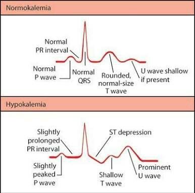
Visual For Ecg Changes For Different Electrolyte Imbalances Nursing School Survival Nursing School Notes Medical School Studying

Components Of The Ecg Strip Emergency Nursing Ecg Rhythms Cardiac Rhythms

T Waves In Ischemia Hyperacute Inverted Negative Wellen S Sign De Winter S Sign With Images Medical School Motivation Acute Coronary Syndrome Percutaneous Coronary Intervention

Medical Examinations Pulmonary Embolism Pulmonary Ekg

Junctional Rhythm Ecg Interpretation Ekg Cardiac Nursing

Ekg Injuries Used To Be Monitor Tech In An Icu Nurse Cardiac Nursing Nursing School

File T Wave Morphology Png Bestand

Ecg Presentation Slide 1 St Segment T Wave Segmentation Interactive Presentation Nursing Study
Pin On Ecg

Normal Ecg From 29 Y O Asymptomatic Athlete Sinus Bradycardia Early Repolarisation With St Elevation Arrows And Pe St Elevation Normal Ecg Cardiac Disease

Pin On Beautiful Body Art

Pin On Cardiac Rhythms

Figure 1 Discordance And Concordance Between Qrs Complex And St T Segment Ecg Interpretation Qrs Complex Normal Ecg

Ecg T Wave Checklist Ecg Interpretation Qrs Complex Interpretation

Junctional Rhythm If The Heart Rate Is Slow 40 55 Min The Qrs Complex Is Normal The P Waves Are Possibly Not Seen Then T Ekg Interpretation Arrythmias Ekg

Ekg Evolution Hyperacute T Waves Immediately 6 24hrs St Segment Elevation Immediately 1 6 Weeks Myocardial Infarction Cardiology Cardiac Nursing

Typical Ecg Abnormalities In Brugada Syndrom Brugada Syndrome St Elevation Abnormal
Click Through To Watch The Full Easy Ekg Interpretation Video Learn Ekg Interpretation In 10 Incredibly Easy Ste Nurse Nursing School Survival Cardiac Nursing

Pin On Medicine

Ekg Changes In Electrolyte Imbalances Nclex Quiz Electrolytes Imbalance Best Nursing Schools Pediatric Nursing

Heart Beat Ecg Tattoo Tattoos And Piercings Tattoos

Ecg Study Cards Sinus Tachycardia Pulmonary Embolism Icu Nursing Pulmonary Embolism Medical Knowledge

Evolution Of Acute Stemi In Order To Diagnose A Stemi One Must First Be Able To Identify The St Segment On The Ekg Cardiology Nursing Emergency Nursing Nurse

Pin By Danielle Sullivan On Medic Pa C Nursing Tips Nurse Cardiac Nursing

Youtube Ekg Interpretation Rhythms Ecg Rhythms

Pin By Shilpa Lakra On Nursing With Images Pr Interval P Wave Qrs Complex

2 646 Likes 5 Comments Medicohub Worldwide Medicohub Worldwide On Instagram Ecg Changes In 2020 Nursing School Survival Electrolytes Nursing Cardiac Nursing

Hypomagnesemia Ekg Various Morphologies Of St T U Abnormalities As Seen In Lead V4 Nurse Nursing Study Paramedic School

Abnormal Ecg Rhythms Medical Surgical Nursing Nursing School Tips Nursing School Notes

Left Atrial Enlargement P Mitrale Right Atrial Enlargement P Pulmonale On Ecg With Images P Wave Ecg Interpretation Qrs Complex

Anterior Myocardial Infarction Ecg Interpretation St Elevation Ecg Rhythms

Left Bundle Branch Block Cardiac Nursing Paramedic Bundle Branch Block

Pin By Jason Winter Ecg Educator On Cardiology Notes Nursing School Notes Nursing Notes Medical Student Study

Related Image Ecg Rhythms Sinusitis Atrial Flutter

Accelerated Junctional Rhythm Ecg Interpretation Ekg Cardiac Nursing

Junctional Escape Beat An Ectopic Junctional Beat That Occurs Late Within An Underlying Rhythm P Wave Will Be Inv P Wave Ekg Interpretation Nursing School Tips

R E B E L Ecg Of The Week Wellens Syndrome Or Stemi

Progression Of An Acute Myocardial Infarction Nurses Tips Nurse Icu Nursing Emergency Nursing

Pin On Ecg

St Segment Elevation In Acute Myocardial Ischemia And Differential Diagnoses Ecg Interpretation St Elevation P Wave

