Inverted T Wave On Ecg Causes
The T Wave Physiology Variants And Ecg Features Ecg Echo
T Wave Litfl Medical Blog Ecg Library Basics
T Waves In Ischemia Hyperacute Inverted Negative Wellen S Sign De Winter S Sign Ecg Echo
T Wave Wikipedia

T Wave Inversion In Leads V1 V6 In A 38 Year Old Symptomatic An Download Scientific Diagram

Causes Of T Wave Inversion In Ecg Cardiology And Ccu Facebook

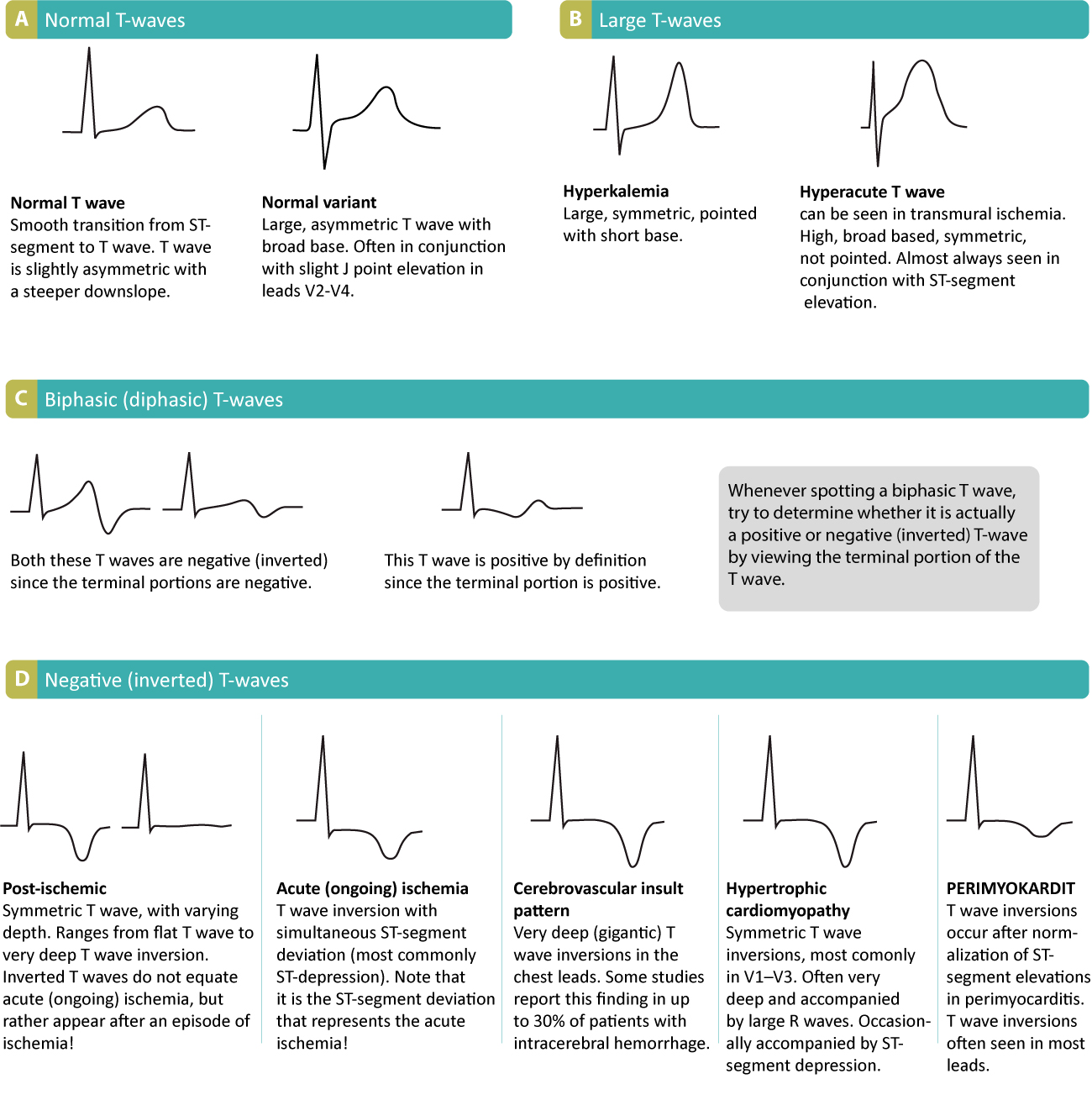
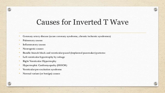
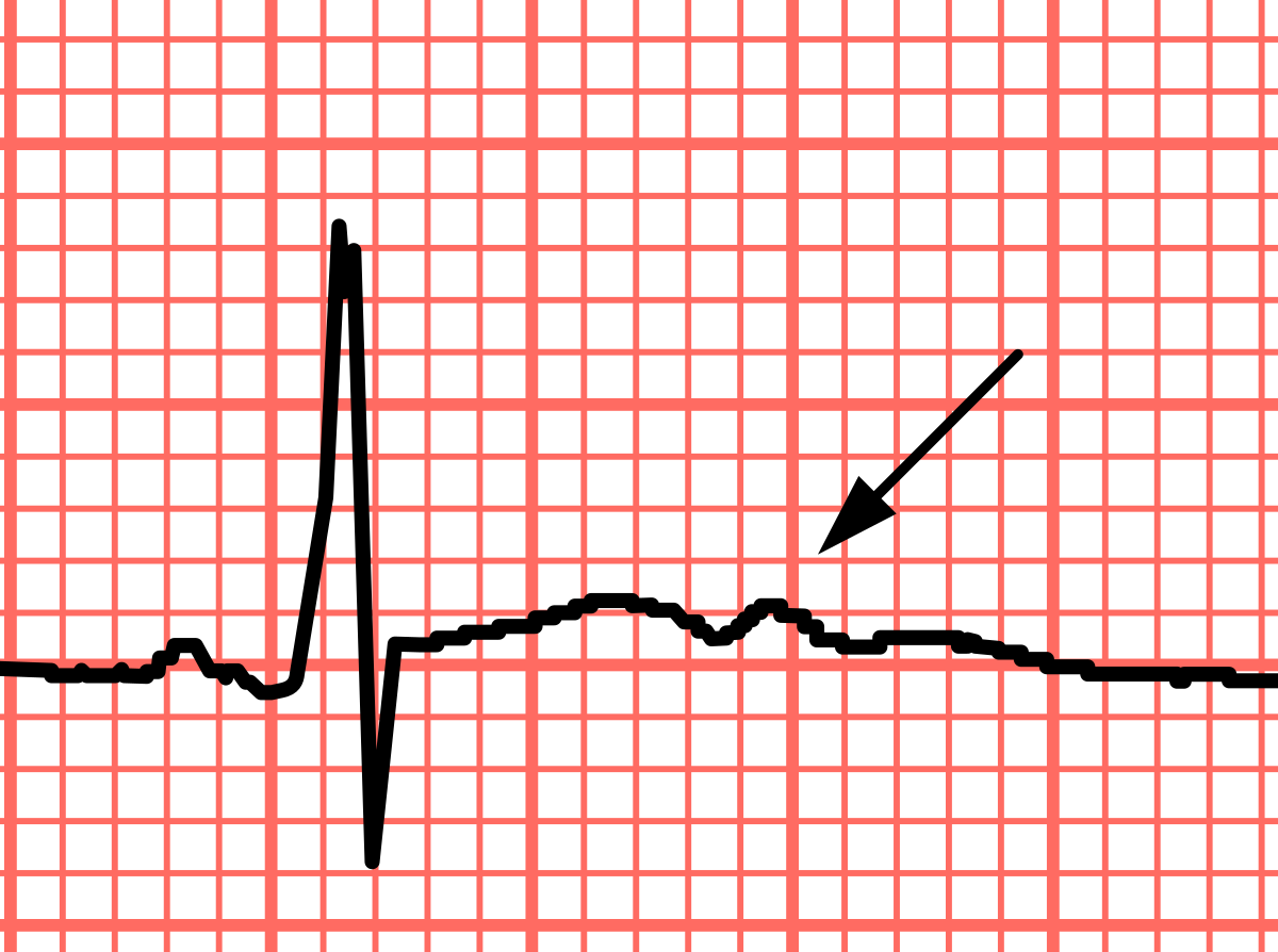
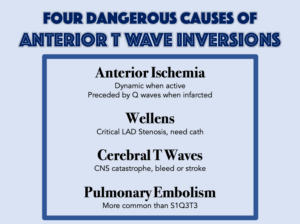
Causes of inverted t waves.
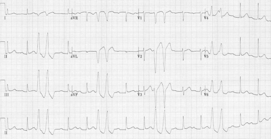
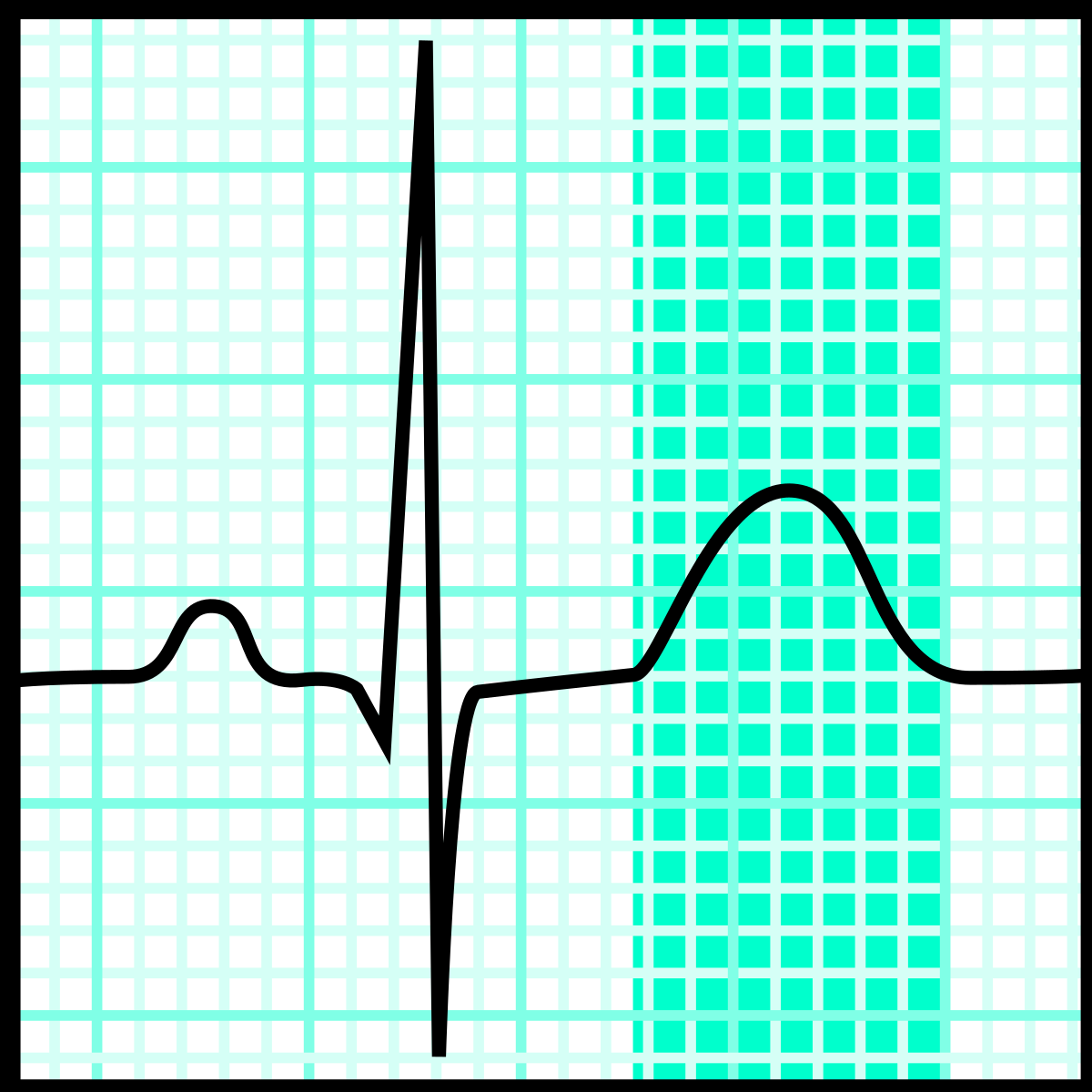
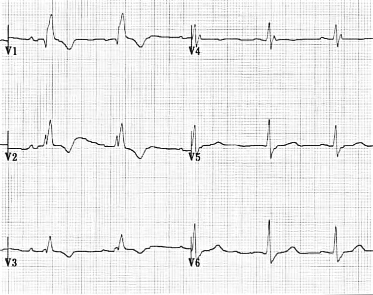
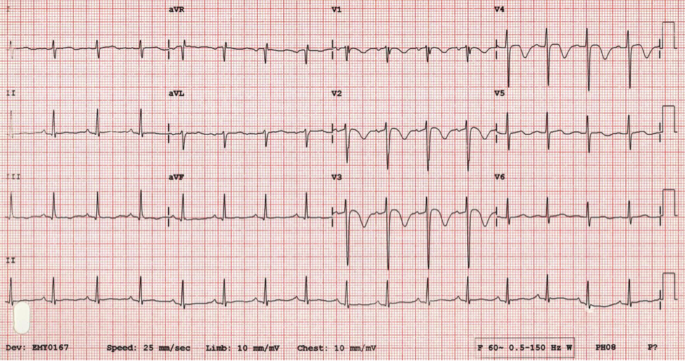
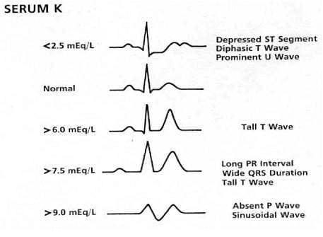
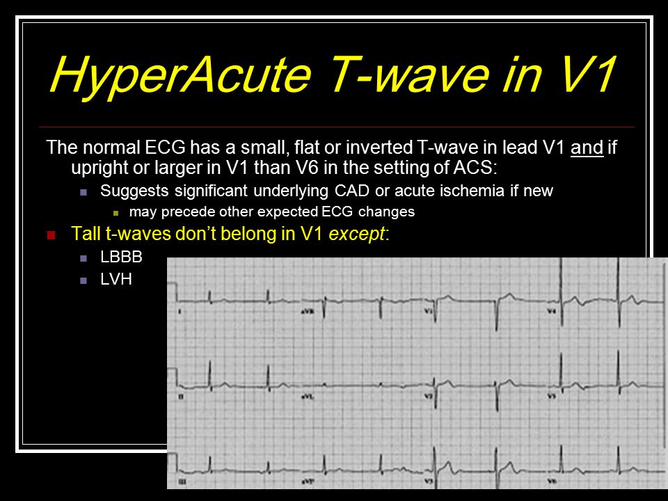
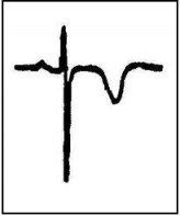
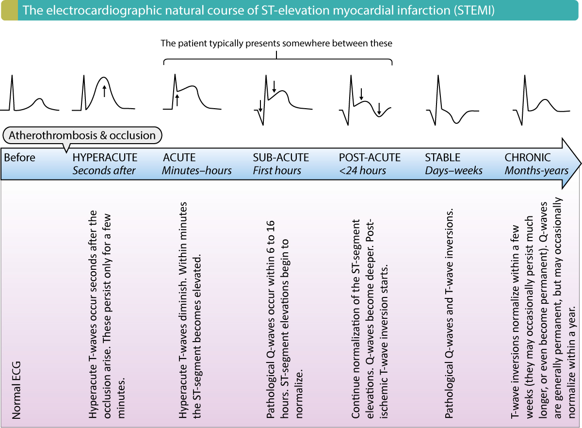
Inverted t wave on ecg causes. The t wave should be concordant with the qrs complex meaning that a net positive qrs complex should be followed by a positive t wave and vice versa figure 17. The t wave is the ecg manifestation of ventricular repolarization of the cardiac electrical cycle. The t wave is normally upright in leads i ii and v3 to v6. Inverted in lead avr.
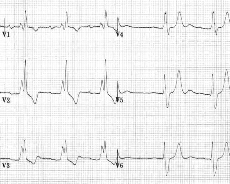
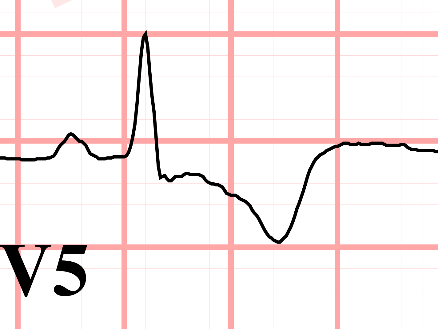
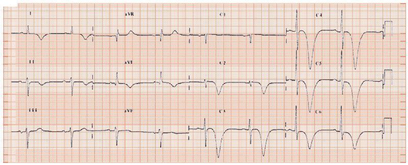
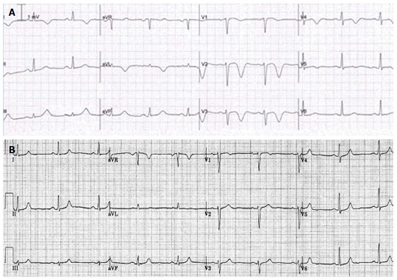
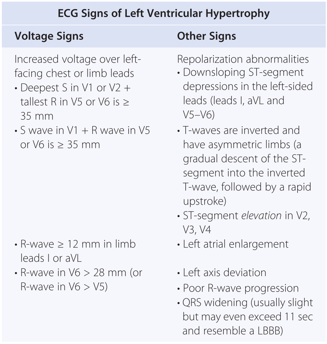
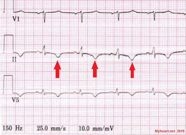
There are several causes which may cause abnormal inversion of t wave. Inverted t waves associated with cardiac signs and symptoms chest pain and cardiac murmur are highly suggestive of myocardial ischaemia. Inverted t waves found in leads other than the v1 to v4 leads is associated with increased cardiac deaths. Inverted t waves mean on an ecg that you should go for further testing.
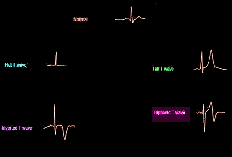
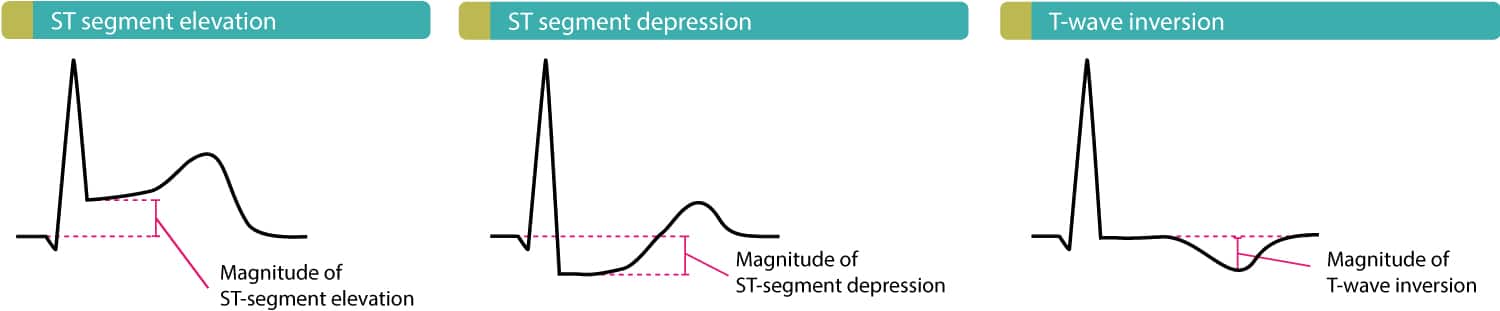
For instance a single inverted t wave in either lead iii or avf can be a normal variant. In normal ecg readings the t wave should be upward. If the readings show different characteristics then you have inverted t waves. Causes of t wave abnormality on ecg.
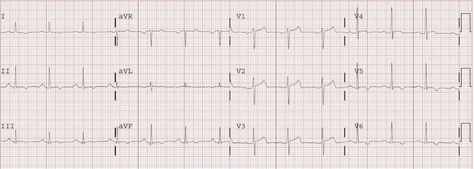
Inverted t wave is considered abnormal if inversion is deeper than 1 0 mm. Otherwise there is discordance opposite directions of qrs and t which might be due to pathology. And variable in leads iii avl avf v1 and v2. Inverted t waves are always noted in the avr and v1 leads.
In general an inverted t wave in a single lead in one anatomic segment ie inferior lateral or anterior is unlikely to represent acute pathology. Here are some of the most common reasons for inverted t waves. The interpretation of the ecg in the context of the individual patient presentation is mandatory. Thus t wave inversions in leads v1 and v2 may be fully normal.
Lesson Title The T Wave
What Is T Wave Inversion Quora
Deep T Wave Inversion Thoracic Key

Mechanism Of Ischemic T Wave Inversion Youtube
Ecg T Wave Inversion Dr Malala Rajapaksha Cardiology Unit Genera

Dr Smith S Ecg Blog Reversible T Wave Inversion It Reverses Then Evolves Then Reverses When Ischemia Is Gone Normalization Of T Waves Not Pseudonormalization

St Depression T Wave Inversion Causes Pathophys Diagnosis Cardiology Medstudent Ekg

Ecg Normal And Inverted T Wave Waves Normal Ekg
Basic Electrocardiography Guide To Diagnostic Tests
U Wave Wikipedia
The Inverted T Wave Differential Diagnosis In The Adult Patient
68 Causes Of T Wave St Segment Abnormalities Learntheheart Com

Chest Pain With Diffuse T Wave Inversion Photo Quiz American Family Physician

Resting 12 Lead Ekg Showing Symmetric T Wave Inversion In Right Download Scientific Diagram
T Wave Inversion Test Findings Medschool

Ecg In A Patient With Arvd C Epsilon Waves And Inverted T Waves Download Scientific Diagram
Cardiac And Non Cardiac Causes Of T Wave Inversion In The Precordial Leads In Adult Subjects A Dutch Case Series And Review Of The Literature
Right Bundle Branch Block Rbbb Litfl Ecg Library Diagnosis
Https Encrypted Tbn0 Gstatic Com Images Q Tbn 3aand9gcrpzmybk 8i8errcymlgjsala2cz97lgve 7noquyscvllzqdr2 Usqp Cau
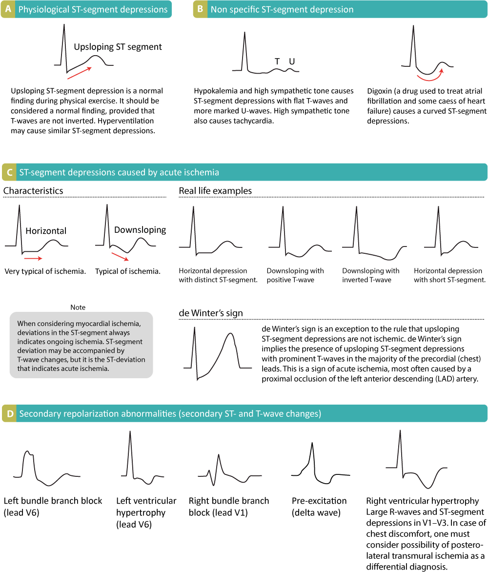
St Segment Depression In Myocardial Ischemia And Differential Diagnoses Ecg Echo
Emdocs Net Emergency Medicine Educationecg Pointers Intracranial Hemorrhage Emdocs Net Emergency Medicine Education

Dr Smith S Ecg Blog Chest Pressure During Exertion Evolution Of Inverted T Waves And Troponins Surprise Angiogram
Ecg T Wave Article Statpearls

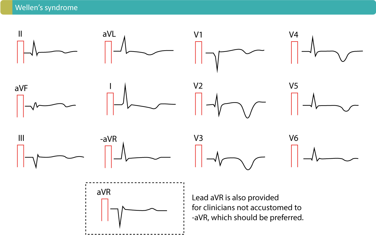
Wellens Syndrome Litfl Medical Blog Ecg Library Eponym
Emdocs Net Emergency Medicine Educationwellens Syndrome Emdocs Net Emergency Medicine Education
T Wave Inversions Maimonides Emergency Medicine Residency

Pdf Cardiac And Non Cardiac Causes Of T Wave Inversion In The Precordial Leads In Adult Subjects A Dutch Case Series And Review Of The Literature

Discordant Ecg Findings In A Man With Chest Pain Consultant360

Ekg Showing Atrial Fibrillation T Wave Inversion At Lead Iii And Download Scientific Diagram
Dr Smith S Ecg Blog 15 Yo Aam With St Elevation And T Wave Inversion Hypertrophic Cardiomyopathy Or Normal Variant
Clinical Application Of Ecg In Chest Pain Acute Myocardial Infarction Ecg Echo
Paediatric Ecg Interpretation Litfl Medical Blog Ecg Library Basics
Top Ten Or 11 Ekg Killers Ppt Video Online Download

Evaluation Of Exercise Stress Test Ecg Symptoms Blood Pressure Heart Rate Performance Ecg Echo
The Inverted T Wave Differential Diagnosis In The Adult Patient Practical Cardiology

Ecg What About U Waves Maimonides Emergency Medicine Residency

Abnormal T Waves Thoracic Key
Confusing Conditions St Segment Depressions And T Wave Inversions Chapter 6 Critical Cases In Electrocardiography
The Ecg In Assessment Of Myocardial Reperfusion Ecg Echo

The 12 Lead Ecg Identified A Normal Sinus Rhythm With Inverted T Waves Download Scientific Diagram
Women And Stress Cardiomyopathy Or Takotsubo Myheart
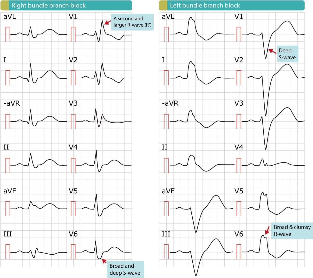
Right Bundle Branch Block Rbbb Ecg Criteria Definitions Causes Treatment Ecg Echo